
Australian Polyclinic · Pleural health guide
Pleural Effusion: Causes, Symptoms, Tests and Treatment
Fluid around a lung is a finding that needs an explanation. Learn why it develops, how doctors investigate it and when drainage or further treatment may help.
Medically reviewed and approved by Dr G Sarwar Chaudhry, MBBS, FRACP (Australia), FCCP (USA), Consultant Pulmonologist and Sleep Physician, on 9 October 2026.
What is a pleural effusion?
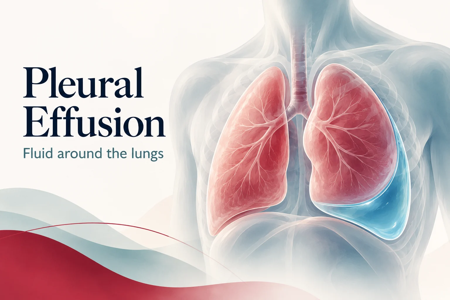
The pleura consists of thin layers covering the lungs and lining the inside of the chest wall. A small amount of fluid normally helps these surfaces slide as you breathe.
A pleural effusion is an abnormal collection of fluid in this space, around the lung rather than inside its air sacs. A larger collection can restrict lung expansion and contribute to breathlessness.
What causes fluid around the lungs?
Several conditions can cause an effusion. The history, imaging and fluid results help distinguish between them.
| Possible cause | What the assessment considers |
|---|---|
| Heart failure | Fluid-pressure changes can produce an effusion, often alongside other signs of fluid overload. |
| Kidney or liver disease | Changes in fluid balance and blood proteins can contribute to pleural fluid. |
| Pneumonia or pleural infection | An effusion may accompany pneumonia. Infected or complicated collections may need drainage as well as antibiotics. |
| Tuberculosis | An important possibility in Pakistan; fluid tests and sometimes pleural tissue are needed to establish the cause. |
| Cancer | Lung cancer and other cancers can cause pleural fluid; imaging and appropriate sampling help assess this. |
| Other conditions | Pulmonary embolism, autoimmune inflammation, chest injury and certain medicines may also be relevant. |
Doctors may describe fluid as transudative or exudative, based on its chemistry and the clinical context. These categories guide investigation but do not identify the exact cause on their own.
2, 3Common symptoms and imaging findings
Possible symptoms include shortness of breath, reduced exercise tolerance, cough and chest pain that worsens with deep breathing. A small effusion may cause no obvious symptoms and be found incidentally.
Fever, weight loss, leg swelling or other associated symptoms can provide clues, but no symptom pattern proves the cause. An unexplained effusion on one side deserves assessment; neither a one-sided nor a two-sided collection is diagnostic by itself.
Bring earlier scans if available. Comparing the size and appearance of the collection over time can help the clinician understand what has changed.
3, 4How is a pleural effusion investigated?
History, examination and imaging
The clinician reviews breathing symptoms, infection history, smoking, previous cancer, occupational exposures, medicines and heart, kidney or liver disease. Oxygen levels and other clinical findings help determine urgency.
A chest X-ray can show fluid. Thoracic ultrasound identifies its position and guides safe sampling or drainage. CT may provide more detail about the lungs and pleura when the cause is uncertain or further investigation is needed.
Blood tests and fluid analysis
Selected blood tests can help assess possible causes. If sampling is indicated, the fluid may be examined for protein and other chemistry, cell counts, infection and cancer cells. Additional tests depend on the clinical question.
Not everyone needs every test or an immediate aspiration. For example, a typical effusion associated with known fluid overload may initially be managed by treating that cause and reviewing the response.
3, 5What happens during pleural fluid aspiration?
Pleural aspiration, also called thoracentesis, removes fluid with a needle or small catheter. A small sample can help diagnose the cause; removing more fluid may relieve breathlessness in selected patients.
Ultrasound is used to identify a suitable site. The procedure is usually performed with local anaesthetic by trained staff in an appropriate clinical setting. Possible complications include discomfort, bleeding, infection and pneumothorax.
Tell the team about blood-thinning medicines, bleeding problems, allergies and previous procedures. Follow the team's instructions; do not stop prescribed anticoagulants yourself.
Suspected pleural TB also requires careful interpretation. A specialist may recommend microbiological tests, selected fluid markers and/or pleural tissue; appearance alone cannot establish TB.
3, 5, 6How is a pleural effusion treated?
Treatment depends on the cause, symptoms, size of the effusion and how the lung responds when fluid is removed.
Treat the underlying condition
Management may include treatment for heart failure or other organ disease, antibiotics for bacterial infection, a full TB treatment programme when TB is confirmed, or cancer-directed treatment.
Drain fluid when appropriate
Symptomatic or infected collections may require aspiration or a chest drain. Small, stable effusions do not always need drainage.
A parapneumonic effusion accompanies pneumonia. Some settle with antibiotics, while complicated infection needs drainage. Empyema means pus within the pleural space and usually requires hospital management.
Selected pleural infections may need medicines delivered through a drain or surgery if initial treatment is insufficient. The plan is reviewed according to clinical progress and imaging.
3, 7Read our pneumonia and recovery guide for information about chest infection and follow-up.
What if the fluid is cancer-related or keeps returning?
A pleural effusion does not automatically mean cancer. When cancer is established as the cause, care usually addresses both the tumour and the fluid-related symptoms.
For recurrent symptomatic malignant effusion, options may include talc pleurodesis, which helps the pleural surfaces adhere, or an indwelling pleural catheter, which allows repeated drainage. Choice depends on whether the lung can re-expand, the person's preferences and practical support.
These procedures relieve or manage fluid; they are not a substitute for discussing appropriate oncology treatment. Our lung cancer guide explains biopsy, staging and modern treatment choices.
3, 8Follow-up and assessment in Lahore
A follow-up plan should explain the working diagnosis, results still awaited, whether imaging needs repeating and who will review a recurrent collection. Seek earlier care if breathing worsens, fever develops or significant chest pain appears.
You can consult Dr G Sarwar Chaudhry at Australian Polyclinic for outpatient respiratory assessment and review of pleural imaging and results. Bring scan images, reports, a medication list and previous fluid or biopsy results.
Ultrasound-guided sampling, chest drainage, pleural biopsy and hospital treatment may need referral to an appropriate procedural or inpatient service. Investigation arrangements are discussed according to your clinical needs.
Frequently asked questions
Is pleural effusion the same as fluid inside the lungs?
No. Pleural effusion is fluid in the space around a lung. Pulmonary oedema involves fluid within lung tissue and air sacs. The two conditions can coexist, but their assessment and treatment differ.
Does fluid around the lung always mean cancer?
No. Heart failure, infection, tuberculosis, kidney or liver disease and other conditions can cause an effusion. The cause is established through clinical assessment and appropriate investigations.
Does every pleural effusion need drainage?
No. Drainage depends on symptoms, the likely cause, size and whether infection or another important diagnosis is suspected. Some small effusions improve when the underlying condition is treated.
Can a negative fluid test rule out cancer?
Not always. Cancer cells are not found in every cancer-related effusion. Further investigation may be needed when imaging or other findings remain concerning.
Why can fluid return after it has been drained?
Fluid can return if the underlying condition is still active. Recurrent collections may need treatment of the cause and, in selected cases, pleurodesis or an indwelling pleural catheter.
Can pleural tuberculosis be diagnosed from the fluid alone?
Sometimes fluid tests support or establish the diagnosis, but they are not always sufficient. Additional microbiology, selected markers or pleural tissue may be needed, depending on the findings.
Related patient guides
Have you been told there is fluid around a lung?
Bring your imaging and reports for a respiratory assessment. Understanding the cause is the first step towards an appropriate treatment plan.
Australian Polyclinic · First Floor, Building 19, CCA, DHA Phase 5, Lahore.
Clinic: Monday–Saturday, 2:00 pm–9:00 pm. Dr Sarwar consultations: 4:00 pm–8:00 pm. Please confirm availability. Sunday closed.
Clinical references and further reading
References checked on 9 October 2026.. Individual care and local availability may differ.
- NHLBI, NIH: Types of pleural disorders—pleural effusion, pleurisy and pneumothorax.
- NHLBI, NIH: Pleural disorders—causes and risk factors.
- British Thoracic Society: Guideline for Pleural Disease, 2023. Investigation of effusion, pleural infection and pleural malignancy.
- NHLBI, NIH: Pleural disorders—symptoms.
- NHLBI, NIH: Pleural disorders—diagnosis.
- British Thoracic Society: Clinical Statement on Pleural Procedures, 2023.
- NHLBI, NIH: Pleural disorders—treatment.
- British Thoracic Society: Quality Standard for Pleural Disease, 2026.
This article provides general patient education and does not replace individual medical assessment. Do not start, stop or change prescribed treatment solely on the basis of this guide. Seek urgent care for severe or worsening symptoms.